Essential MRI Brain Anatomy for Reporting: A Radiologist’s Quick Reference
Essential MRI Brain Anatomy for Reporting: A Radiologist’s Quick Reference
A solid grasp of neuroanatomy is critical for accurate and confident MRI brain reporting. This article serves as a quick visual and descriptive reference for the key anatomical structures you’ll encounter daily. Perfect for residents needing a refresher or any radiologist wanting to sharpen their skills for clearer, more precise reports.
Understanding MRI brain anatomy for reporting is crucial for radiologists aiming to deliver accurate and confident interpretations. This guide provides a comprehensive overview of the essential anatomical structures encountered in daily practice, enhancing the clarity and precision of radiological reports.
Key Anatomical Structures in MRI Brain Anatomy for Reporting
For effective MRI brain reporting, familiarity with key anatomical structures is essential. These include:
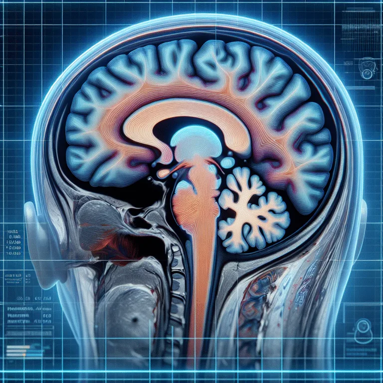
- Cerebral Hemispheres: Comprising the frontal, parietal, temporal, and occipital lobes, each with distinct functions and clinical significance.
- Basal Ganglia: Involved in movement regulation, often evaluated in cases of movement disorders.
- Thalamus: Acts as a relay station for sensory and motor signals.
- Brainstem: Includes the midbrain, pons, and medulla, crucial for autonomic functions.
- Cerebellum: Responsible for coordination and balance.
Recognizing these structures on MRI scans is vital for accurate diagnosis and reporting. For instance, the basal ganglia’s role in movement disorders makes it a frequent subject of study in cases of Parkinson’s disease.
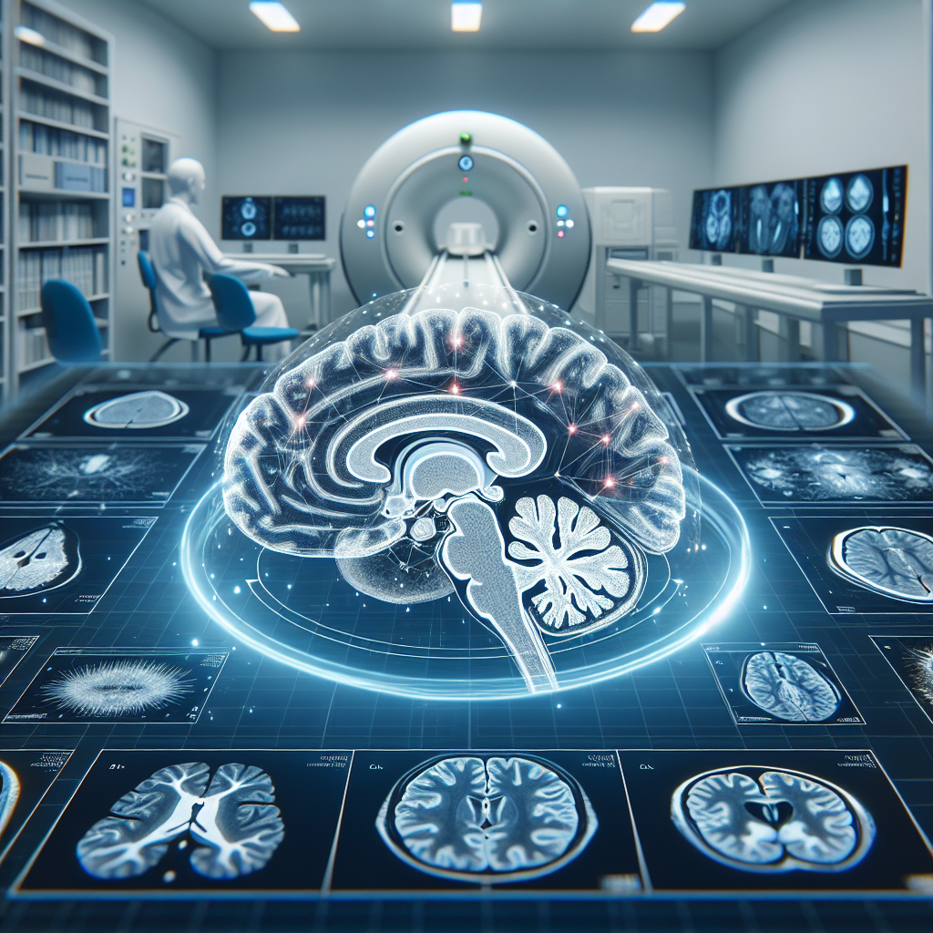
Understanding MRI Sequences in Brain Anatomy Reporting
Different MRI sequences provide varied insights into brain anatomy. Common sequences include:
- T1-weighted images: Useful for evaluating normal anatomy and identifying fat or hemorrhage.
- T2-weighted images: Highlight fluid-filled structures and are instrumental in detecting edema or lesions.
- FLAIR (Fluid-Attenuated Inversion Recovery): Suppresses fluids to better visualize lesions adjacent to CSF spaces.
- DWI (Diffusion-Weighted Imaging): Critical for identifying acute ischemic strokes.
Each sequence provides unique information, contributing to a comprehensive understanding of the brain’s anatomy and pathology. For example, Radiopaedia offers detailed insights into the applications of these sequences in clinical practice.
Sample Radiology Report Template for MRI Brain
Creating a structured and clear report is essential for effective communication. Below is a brief template snippet for an MRI brain report:
MRI Brain Report:
- Date: [Insert Date]
- Patient ID: [Insert ID]
- Indication: [Insert Clinical Indication]
- Technique: [List MRI sequences used]
- Findings:
  - Normal anatomy: [Describe normal structures]
  - Abnormal findings: [Detail any lesions or anomalies]
- Impression: [Summarize key findings and potential diagnoses]
Using a consistent template ensures all necessary details are included, facilitating better understanding and decision-making by referring physicians.
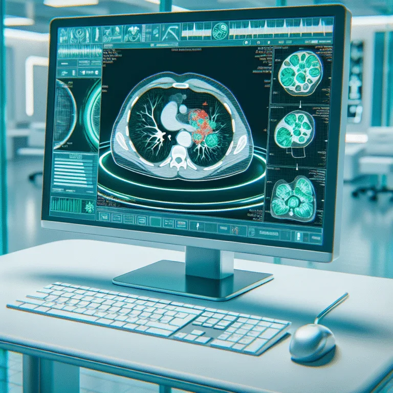
Enhancing Report Accuracy with AI Assistance
Integrating AI tools can significantly enhance the accuracy and efficiency of MRI brain reporting. AI algorithms can assist in identifying subtle abnormalities and ensuring comprehensive coverage of all anatomical structures. For instance, recent studies suggest that AI can improve diagnostic accuracy in neuroimaging.
Adopting AI solutions like Rad Report AI can streamline the reporting process, allowing radiologists to focus on clinical insights and patient care.
Conclusion
A thorough understanding of MRI brain anatomy for reporting is indispensable for radiologists. By mastering key anatomical structures and utilizing advanced imaging sequences, radiologists can enhance the accuracy and clarity of their reports. Incorporating AI tools further optimizes the reporting process, ensuring precise and efficient delivery of diagnostic information.
Embrace the potential of AI in radiology to enhance your reporting capabilities and provide superior patient care.
🚀 Try Rad Report AI For Free — and experience faster, smarter reporting today.