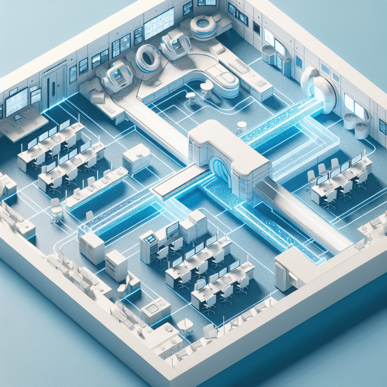
Improving Clarity in CT Scan Reports for Clinicians: 5 Simple Tweaks
Improving Clarity in CT Scan Reports for Clinicians: 5 Simple Tweaks
A report that isn’t clearly understood can delay patient care. Sometimes, small changes can make a huge difference. This article offers five simple but powerful tweaks you can apply to your CT scan reports to improve clarity, eliminate ambiguity, and ensure your referring clinicians can act on your findings with confidence.
Improving clarity in CT scan reports for clinicians is crucial for effective communication and timely patient care. Radiologists often face the challenge of ensuring that their findings are clearly understood by referring clinicians. By implementing a few strategic changes, radiologists can enhance the clarity of their reports, thus facilitating better clinical decision-making.
1. Use Structured Reporting Formats
Structured reporting formats can significantly enhance clarity by providing a consistent framework for presenting findings. This approach reduces ambiguity and ensures that all critical information is included. Structured reports typically follow a standardized template that includes sections such as:
- Patient Information: Name, age, and relevant clinical history.
- Study Details: Type of scan, date, and technical parameters.
- Findings: Detailed observations with precise anatomical descriptions.
- Impressions: Summary of key findings and potential diagnoses.
Adopting structured reporting can lead to more efficient communication and reduce the likelihood of misinterpretation. For more on structured reporting, consider exploring resources from the Radiological Society of North America.
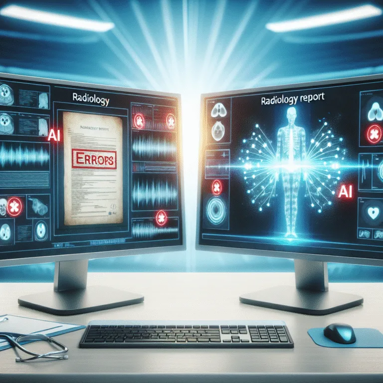
2. Implement Standardized Terminology
Using standardized terminology is another effective way to improve clarity. Consistent language helps avoid confusion and ensures that all clinicians interpret the findings in the same way. The use of standardized terms, such as those found in the American College of Radiology’s lexicon, can be particularly beneficial.
For example, instead of using vague terms like “lesion,” specify the type, size, and location. This specificity helps clinicians understand the clinical significance of the findings more clearly.
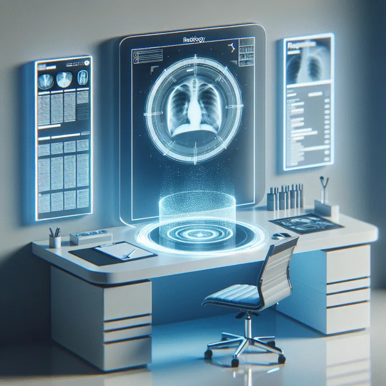
3. Enhance Visual Aids
Visual aids, such as annotated images and diagrams, can greatly enhance the clarity of CT scan reports. By highlighting key findings directly on the images, radiologists can provide a visual reference that complements the written report. This approach can be particularly useful for complex cases where verbal descriptions alone may not suffice.
Ensure that images are of high quality and that annotations are clear and concise. Tools like Rad Report AI can assist in efficiently integrating annotated images into reports.
4. Provide Clear and Concise Impressions
The impression section of a CT scan report is often the most critical for referring clinicians. It should succinctly summarize the key findings and their potential clinical implications. Avoid unnecessary jargon and focus on delivering a clear, concise message.
Consider using bullet points or numbered lists to highlight important points. This format can make it easier for clinicians to quickly grasp the essential information.
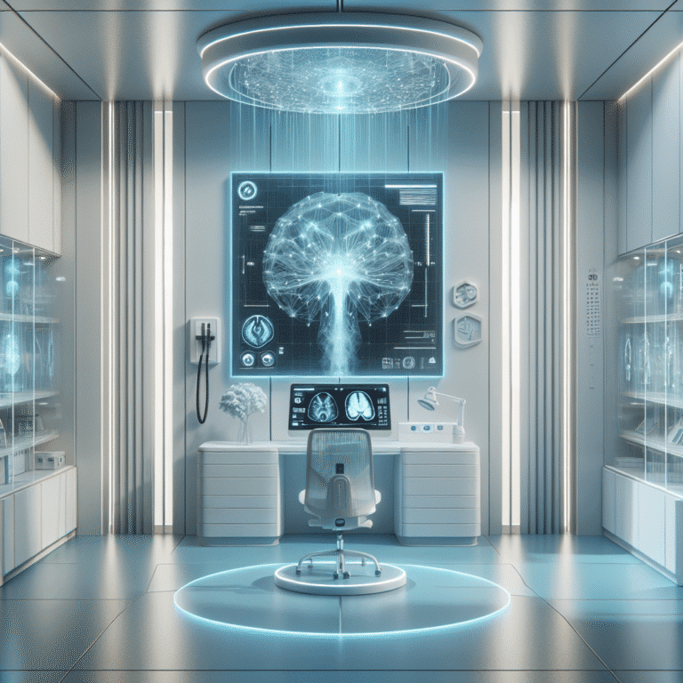
5. Leverage AI and Voice Dictation
AI and voice dictation technologies can play a pivotal role in improving the clarity of CT scan reports. These tools can help radiologists streamline the reporting process, reduce errors, and enhance the overall quality of the report.
For instance, AI algorithms can assist in identifying and highlighting key findings, while voice dictation can speed up the documentation process. By integrating these technologies, radiologists can focus more on analysis and less on manual data entry.
Sample Radiology Report Template
Below is a sample snippet of a structured radiology report template:
Patient Information:
- Name: John Doe
- Age: 45
- Clinical History: Persistent cough, smoker

Study Details:
- Type: Chest CT
- Date: 2023-10-15
- Technique: Axial slices, contrast-enhanced

Findings:
- Right upper lobe mass measuring 3 cm
- No lymphadenopathy observed

Impressions:
- Suspicious for primary lung carcinoma
- Recommend biopsy for further evaluation
By implementing these five simple tweaks, radiologists can significantly improve the clarity of their CT scan reports, leading to better communication and enhanced patient care. As the field of radiology continues to evolve, leveraging technology and standardized practices will be key to maintaining high-quality reporting.
🚀 Try Rad Report AI For Free — and experience faster, smarter reporting today.